Catabolism Pathways
Written by Ben Bunting: BA, PGCert. (Sport & Exercise Nutrition) // British Army Physical Training Instructor // S&C Coach.
--
Question: What are catabolic pathways?
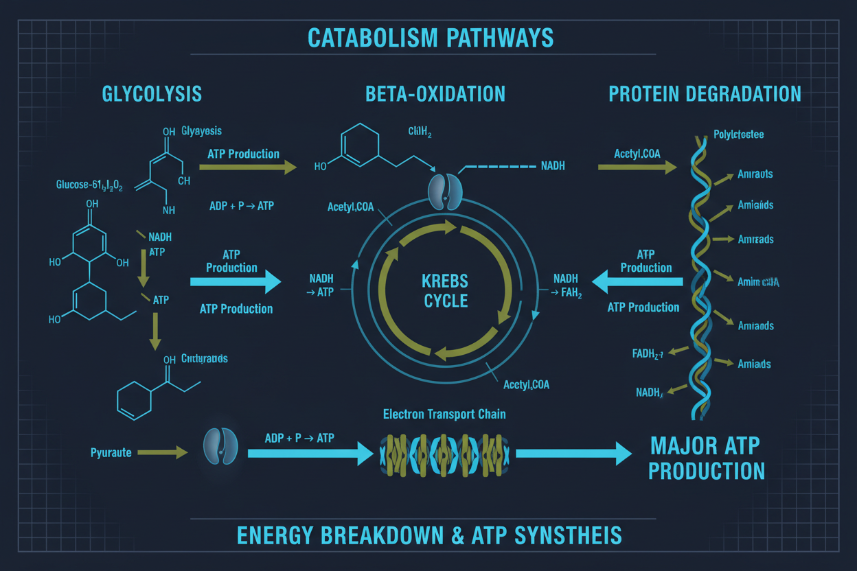
Answer: Catabolic pathways are metabolic pathways that break down complex molecules into simpler ones, releasing energy in the process.
These pathways break down carbohydrates, fats, and proteins to produce energy for cellular processes.
An Overview of Catabolic Pathways
In this article, we will take a closer look at metabolic pathways - linked chemical reactions that produce something and transform it into another substance.
These pathways consist of linked reactions which feed off each other, taking in products of previous reactions and turning them into something new.
These chemical reactions start with starting molecules and turn them into end products through various catabolic (degradative) or anabolic (building up) processes.
Catabolic reactions break down large molecules into smaller ones; anabolic reactions build larger ones from simpler components.
All biological reactions require and release energy; many of them can be catalysed by proteins called enzymes.
Breakdown energy is converted to Adenosine Triphosphate, or ATP, the energy currency of cells. This energy can then be used for anabolic processes, stored as fat or nucleic acids, or used to power anaerobic metabolism.
Most organisms capture energy from the environment to power cellular functions, most commonly through catabolic reactions such as cellular respiration, which breaks down glucose to produce energy for cells.
Glycolysis produces pyruvate that can enter either the citric acid cycle or oxidative phosphorylation pathways. Alternatively, it can be routed through the pentose phosphate pathway to produce five-carbon sugars needed for DNA and RNA synthesis.
Furthermore, this pathway produces acetyl-CoA, which can then be converted to ATP via mitochondrial oxidative phosphorylation.
Catabolic Pathways - Breaking Down Molecules for Energy
Organisms need energy for daily functioning. Organisms harvest potential energy stored in carbon and oxygen molecules (CO2 and H2O), harness it, and create ATP via cellular respiration.
One key step involves breaking down sugar molecules into VFAs and CO2. Glucose is one such source that plants can use directly or that bacteria can consume and convert to energy via fermentation.
Both processes involve metabolic pathways - interlinked chemical processes that feed off each other before culminating in production of an end product - something glucose plays a significant role.
Cellular metabolism can be divided into catabolic and anabolic pathways. Catabolic pathways involve degradative chemical reactions that break complex molecules down into smaller ones; examples include glycolysis, the citric acid cycle, and neurotransmitter deamination via oxidative deamination.
These pathways supply both energy and building blocks that will eventually be used to assemble larger macromolecules through anabolic reactions.
For instance, breaking down glucose via glycolysis produces two molecules of ATP that can then be used in biosynthesis to form monosaccharides, nucleotides or amino acids.
Feedback inhibition connects various steps of these pathways. For instance, phosphofructokinase catalyses a reaction in the citric acid cycle that produces more ATP than it needs; this energy is then recycled through subsequent reactions in its cycle.
The Role of IGFs in Catabolism
Catabolic processes entail disassembling complex organic molecules into smaller components. Within the body, this usually means breaking down polysaccharides such as starch and glycogen into monosaccharides for energy use; proteins into amino acids; and nucleic acids into nucleotides.
IGF1 plays multiple roles in metabolism. Recent studies have demonstrated that its deficiency increases insulin resistance, impairs lipid and glucose metabolism and increases oxidative stress in tissues.
Unfortunately, its role as an anabolic factor is further complicated because hepatocytes serve as both sources of IGF1 and receptors for it.
Protein Synthesis
Proteins are among the most abundant macromolecules in organisms, serving numerous essential roles, including structural support, transport, and regulatory processes.
Their production involves several steps involving transcription and translation. During transcription, genetic information is copied into messenger RNA and exported from the nucleus to the cytoplasm, where it attaches to a protein-synthesis machine.
This is called a ribosome, where translation produces a chain of amino acids that later folds into specific protein structures; this complex biological event must be carefully managed by specialists.
Proteins are composed of amino acid monomers produced in cells by synthesising glucose or other carbon sources into amino acid monomers.
Enzymes then combine them to form polypeptide chains, which fold into secondary, tertiary, and quaternary structures before being transported throughout cells to fulfil their roles.
A significant amount of metabolic energy consumed by cells goes toward maintaining this process - up to 25% according to some estimates!
Previous studies suggest severe burns can induce a catabolic state characterised by muscle wasting and decreased net protein synthesis.
Nutritional support and pharmaceutical interventions with insulin-like growth factor (IGF) or IGFBP-3 have been found to attenuate this muscle catabolism; however, using these agents in humans may produce unwanted side effects.
One study assessed the effect of combining human IGF-1/IGFBP-3 on skeletal muscle metabolism in severely burned children.
Researchers administered IGF-1/IGFBP-3 to 29 patients at 0, 0.5, 1, 2, or 4 mg/kg/day and assessed net protein balance and fractional synthetic rates before and after treatment.
It improved net protein synthesis but did not significantly alter glucose metabolism or plasma urea levels; these changes did not affect gluconeogenesis.
High protein intakes have long been linked to decreased cancer rates and mortality; however, the mechanism remains unexplored.
We have previously shown how the GHR-IGF-1 and mTOR pathways may contribute to the longevity benefits associated with eating more protein.
However, in a recent survey of middle-aged people, it was discovered that even though higher protein consumption reduces risks of cancer and death, it actually increases both all-cause mortality as well as cause-specific deaths.
Lipid Metabolism
Insulin and IGF-1 play an intricate role in glucose and lipid metabolism by acting in concert to produce an integrated response. Of the genes IGF-1 regulates directly, those related to metabolic enzymes are particularly affected.
Partial IGF-1 deficiency reduces hepatic expression of these enzymes, deregulating glucose and lipid metabolism and contributing to metabolic syndrome development.
Blood serum values for glucose, triglycerides and cholesterol, as well as the level of MDA, were examined in untreated Hz mice as well as CO and Hz + IGF-1 animals.
Untreated Hz mice demonstrated significant increases in triglycerides and cholesterol as well as significant decreases in HDL levels compared to CO animals.
With substitution of IGF-1, these parameters returned to similar levels found in CO animals.
IGF-1 also helped normalise expression levels of three enzymes involved in hyperlipidemia, such as g6pc (glucose-6-phosphatase catalytic), Pdk1 (phosphoenolpyruvate carboxykinase 1, cytosolic), and ACLY (ATP citrate lyase), thereby decreasing hyperlipidemia, hyperglycemia, and peroxidative liver damage caused by free radicals.
Reducing expression of acetyl-CoA acetyltransferase 1 (acetyl-CoA acetyltransferase 1), another hepatic enzyme involved in lipid metabolism, was also observed in Hz mice compared with CO animals.
This finding was confirmed by microarray analysis and RT-qPCR, which showed the same trend.
Substitution with IGF-1 increased hepatic expression, suggesting that low circulating IGF-1 levels in Hz mice were responsible for deregulation of lipid metabolism.
IGF-1 levels decline with age, which has been linked with increases in dyslipidemia (cholesterol and triglycerides) as well as hyperglycemia and insulin resistance, resulting in liver damage and mitochondrial dysfunction.
The research indicates that replacing IGF-1 with exogenous IGF-1 at very low doses restores normal levels of these parameters, thereby decreasing dyslipidemia and hyperglycemia and reducing oxidative liver damage. Suggesting this approach as a viable therapeutic strategy for MetS.
Carbohydrate Metabolism
Carbohydrate metabolism refers to a series of biochemical pathways that break down fuel molecules into energy-rich molecules such as ATP, GTP and reduced nicotinamide adenine dinucleotide phosphate (NADH2).
Or reduced flavin adenine dinucleotide (FAD) and reduced nicotinamide adenine dinucleotide phosphate (NADP+).
These molecules can be produced through glycolysis, the citric acid cycle, or the pentose phosphate pathway; mammals regulate carbohydrate metabolism via insulin and glucagon.
Insulin is a polypeptide hormone composed of two chains linked by disulfide bonds. Released by the pancreas in response to food consumption, insulin stimulates glycolysis by binding to its receptors on cells.
Once activated, insulin increases glucose transport into muscle cells and adipose tissue while stopping protein degradation to promote protein synthesis.
Glycogenesis, or the production of glycogen in liver and muscle, is controlled by insulin.
Not all glucose that enters our bodies can be effectively processed. Any excess is stored as glycogen in muscle and adipose tissues for later release during fasting or exercise, or broken down to produce glucose via gluconeogenesis and citric acid cycle reactions stimulated by insulin or growth hormone.
In turn, glucagon or epinephrine inhibits these reactions.
Insulin/glucagon regulation of carb metabolism is complex. When blood sugar levels are elevated, insulin secreted from β cells promotes glycolysis to lower glucose concentration in most cells of the body while inhibiting gluconeogenesis and glycogen breakdown in the liver.
Conversely, when blood sugar levels decrease, glucagon, produced by A cells, triggers glucose production in the liver and muscle and releases it back into circulation by breaking down glycogen in the liver and releasing more glucose into circulation.
Carbs serve a crucial signalling role in cells by attaching to proteins through N-glycosidic bonds and forming glycoproteins, oligosaccharides, or glycopeptides.
Posttranslationally, these glycoproteins can be modified by adding glucosamine, galactose, or N-acetylgalactosamine residues in the endoplasmic reticulum and Golgi apparatus, altering their structural integrity, activity, and stability.
Fat Metabolism
IGFs can stimulate cell and tissue growth in muscles and bones and play an essential role in regulating fat metabolism. This includes stimulating lipid synthesis, increasing transport to mitochondria, and switching over to glycolytic metabolism to help build muscle mass.
IGFs exert their growth-promoting effects regardless of their serum concentration.
However, their actions can be adjusted by binding to proteins known as IGFBPs.
This family has six members that vary in their ability to bind IGFs and regulate their activities; most extracellular IGFs are bound with either IGFBP-3 or -4 in ternary complexes that increase availability by protecting them from degradation and helping them reach receptors more quickly.
These compounds can bind and inhibit IGF receptors, interfering with their actions.
Alternatively, they may promote cell transformation and tumour growth. For instance, IGF-1 helps promote lamellipodia formation, an indicator of cancer cells linked to increased migration and metastasis.
IGF-1 may promote type 1 diabetes in mice when its effects cause their immune systems to attack beta cells of the pancreas that produce insulin, though studies have demonstrated that when levels of IGF-1 are kept at lower levels, it promotes longevity, reduces oxidative stress, and atherosclerosis progression is lessened.
IGF-1 is an essential hormone in our bodies, but its levels may become harmful when they rise too far.
Therefore, eating a well-balanced diet to maintain steady IGF-1 levels is key; intense/strenuous exercise may also help with this goal, although as soon as your body adjusts, it may start decreasing it again - for this reason alone, it is advised not to take IGF-1 supplements without first consulting your physician, especially if you have prediabetes or diabetes.
Conclusion
IGFs circulate in extracellular fluid bound to binding proteins called IGFBPs that act classically to limit access of IGFs to their receptors and modulate IGF actions.
Furthermore, genetically modified animals provide new insights into how these IGFBPs interact with IGFs to regulate growth and metabolism.
IGF system is complex and regulates multiple cellular processes in an endocrine, autocrine and paracrine fashion.
It plays a key role in cell growth, differentiation and metabolism, while many of its effects overlap with those of insulin/INSR.
IGF-1 is a key anabolic hormone that promotes protein synthesis in skeletal muscle during prolonged fasting or after high-protein meals, likely by binding IGFBP-2 as a delivery and linkage molecule, increasing its affinity for the IGF-receptor complex and providing easier access to receptor sites on target cells.
IGF-1 acts to inhibit both hepatic glucose production and pituitary GH secretion in humans and mice.
Furthermore, its actions on muscle free fatty acid metabolism and renal gluconeogenesis appear to play an integral part in increasing insulin sensitivity in type 2 diabetes, although its exact role in increasing insulin sensitivity remains uncertain.